
Single Case Experience of Immunosuppressant Administration to SystemicSclerosis-ILD Patients with Aspergilloma
Article Sidebar
Main Article Content
Abstract
Background: Systemic sclerosis (SSc) is a chronic autoimmune disease that
still poses a great challenge to clinicians. SSc is characterized by immune
dysregulation and progressive fibrosis that typically affects variable internal
organ involvement such as lungs. Interstitial lung disease (ILD) is a common
manifestation of SSc and a leading cause of death. The immunosuppressive
drug is the main treatment to suppress the inflammation process in SSc.
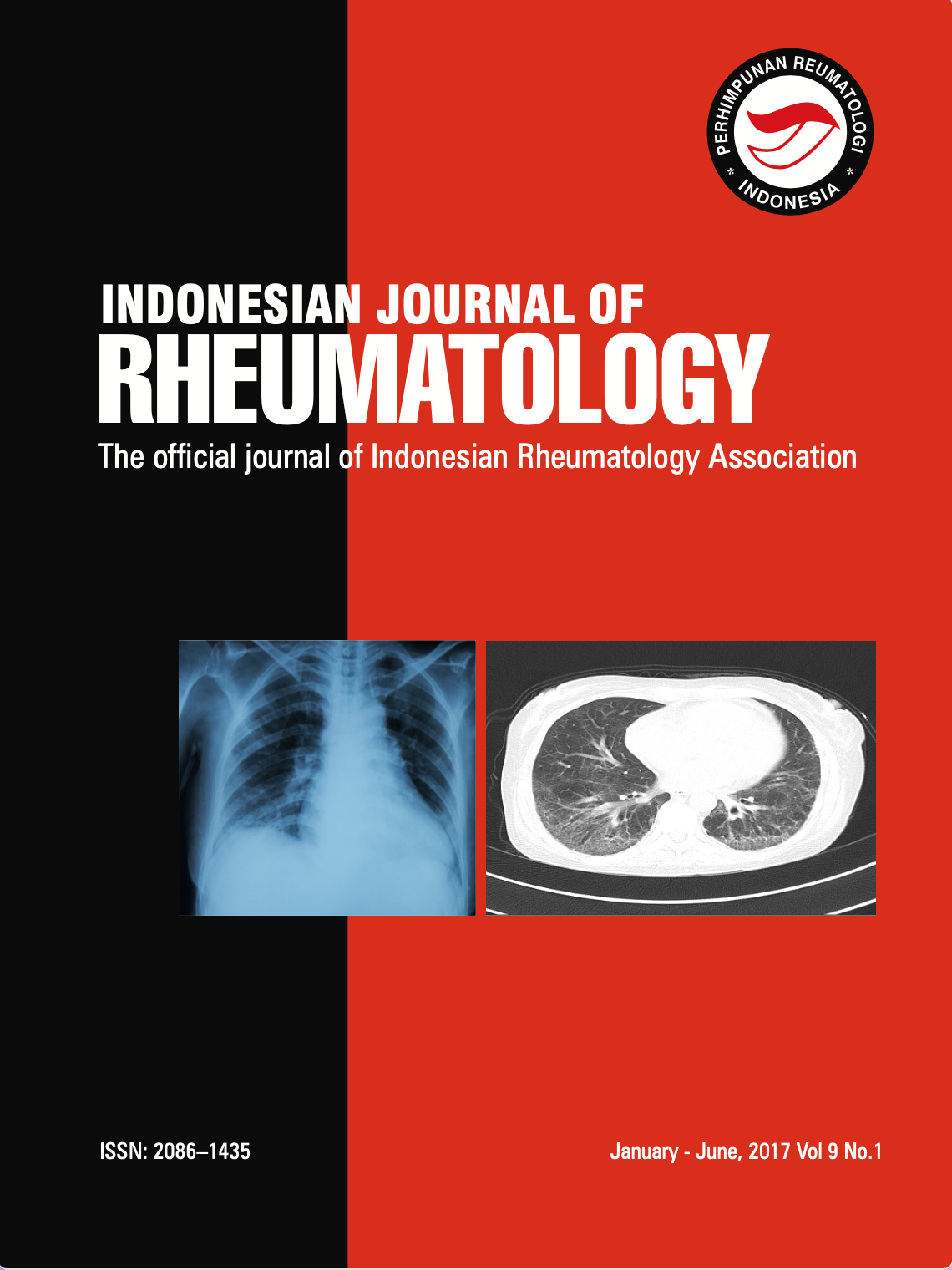
Case presentation: In this case we report a 40-year-old female to suffer ILDSSc.
According to High-Resolution Computed Tomography (HRCT) thorax,
we found that it was interstitial lung disease with aspergilloma. She got
methylprednisolone 3x8 mg and azathioprine 2x50 mg. At the end of the
treatment, the patient showed improvement in her clinical condition and
showed no worsening condition in the HRCT evaluation for her fungal
aspergilloma. Conclusion: Systemic sclerosis (SSc) is a rare autoimmune
disease involving the skin and internal organs. The immunosuppressive
agent is still the drug of choice for most autoimmune diseases.
Immunosuppressive may promote fungal growth and have been associated
with increased risk in most serious fungal diseases including aspergilloma.